Ligament Laxity
Understanding Spinal Ligament Laxity
Ligament Laxity Explained
When Ligaments Fail, Instability Follows
Spinal ligament injuries often go undetected on static imaging like MRI or plain X-rays, yet they can be a major source of chronic pain and long-term impairment. The most affected structures include the posterior longitudinal ligament (PLL), anterior longitudinal ligament (ALL), and alar/accessory ligaments. When these ligaments are compromised, they allow abnormal motion, such as excessive vertebral translation, disc space widening, or C1-C2 instability, which signals functional spinal instability. DXD captures and quantifies these motion patterns to reveal ligament damage that conventional scans miss.
Why Motion Matters
Posterior Longitudinal Ligament
The integrity of the posterior longitudinal ligament (PLL) is demonstrated by a forward (anterior) movement (translational motion) of one vertebrae over the vertebrae below or by the posterior widening of the intervertebral disc space (increased disc angle or angular motion). By measuring these discrepancies of George’s Line (Yochum & Rowe, p. 149), AOMSI can be quantified and correlated with the AMA guides. (5th edition, p. 378–379).
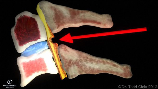
Widening of the posterior disc
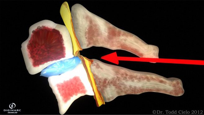
Anterolisthesis
Anterior Longitudinal Ligament
The integrity of the anterior longitudinal ligament (ALL) is demonstrated by a backward (posterior) movement (translational motion) of one vertebrae over the vertebrae below or by the anterior widening of the intervertebral disc space (increased disc angle or angular motion). By measuring these discrepancies of George’s Line (Yochum & Rowe, p. 149), AOMSI can be quantified and correlated with the AMA guides (5th edition, p. 378–379).
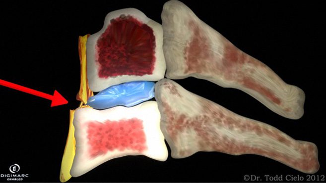
Retrolisthesis
Widening of the anterior disc
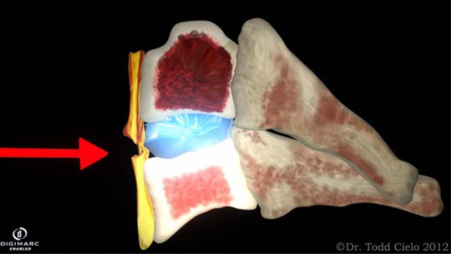
Alar/Accessory Ligaments
The integrity of the posterior longitudinal ligament (PLL) is demonstrated by a forward (anterior) movement (translational motion) of one vertebrae over the vertebrae below or by the posterior widening of the intervertebral disc space (increased disc angle or angular motion). By measuring these discrepancies of George’s Line (Yochum & Rowe, p. 149), AOMSI can be quantified and correlated with the AMA guides. (5th edition, p. 378–379).
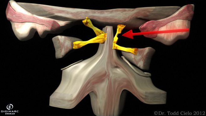
C1 Lateral mass overhang
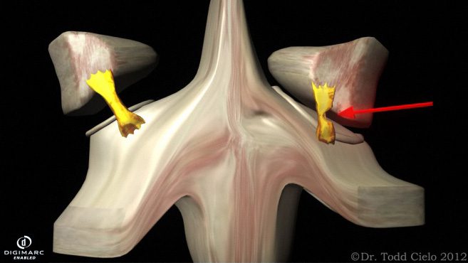
Change in Para-odontoid space
Who DXD Helps
Trusted by Doctors, Attorneys, and Patients
DXD Xray transforms motion X-rays into AMA-compliant ligament injury reports that help doctors diagnose with confidence, attorneys validate or refute claims with objective data, and patients understand what static films may have missed.

For Doctors
DXD provides AMA-compliant injury reports that improve documentation, support clinical decisions, and strengthen reimbursement claims.

For Attorneys
DXD provides objective ligament injury reports that clarify diagnosis, support case strategy, and strengthen personal injury claim outcomes.

For Patients
DXD helps uncover injuries traditional imaging can miss, giving patients the objective proof they need to get answers and better care.

Meet the Founder
About Dr. Cielo
Dr. Todd Cielo is the founder of DXD and a practicing chiropractor with over 20 years of experience in spinal injury care, education, and interdisciplinary collaboration. He is a lecturer for continuing education programs across Florida and the accredited CLE speaker for the Florida Bar Association. Appointed by the Governor to the Florida Board of Chiropractic Medicine, Dr. Cielo brings deep clinical and medico-legal insight to every DXD report. His mission is simple: to bring objectivity, credibility, and better outcomes to soft-tissue injury diagnosis.
Get clarity from the motion experts.
Connect with the DXD team to learn how our ligament analysis can support your case, your care, or your clinic.
Contact Us
Let’s Talk Ligaments…
Got questions about our reports, workflow, or if DXD is right for you? Reach out and we’ll respond within 1 business day.

Email: [email protected]